Lung Parenchyma : Lung Parenchyma Keyword Search Science Photo Library : Pulmonary interstitium is a collection of support tissues within the lung that includes the alveolar epithelium, pulmonary capillary endothelium, basement membrane, perivascular and perilymphatic tissues.
Lung Parenchyma : Lung Parenchyma Keyword Search Science Photo Library : Pulmonary interstitium is a collection of support tissues within the lung that includes the alveolar epithelium, pulmonary capillary endothelium, basement membrane, perivascular and perilymphatic tissues.. He pulmonary parenchyma is the portion of the lung involved in the hematosis or gas transfer. 50% off with $15/month membership. Lung nodules are usually about 0.2 inch (5 millimeters) to 1.2 inches (30 millimeters) in size. Holes in the lungs, known as bullae, can. Lung nodules — small masses of tissue in the lung — are quite common.
It includes the alveolar walls as well as the blood vessels and the bronchi. These components are not homogeneously distributed over the lung and the relative proportion is continuously changing in function of normal physiological events. Talk to a doctor now. Some definitions also include other structures and tissues within the lung parenchyma. The lung parenchyma is that portion of the lungs involved in gas exchange.
Medicohub Worldwide 1m On Instagram A Pulmonary Alveolus Is A Hollow Cup Shaped Cavity Found In The Lung Parenchyma Where Gas Exchange Takes Place Lung Alveo I 2021 from i.pinimg.com However, some authors include other structures and tissues within the definition. What is a soft tissue pulmonary nodule in lung. The alveoli are held open by the transpulmonary pressure, or prestress, which is balanced by tissues forces and alveolar surface film forces. In the original analysis, the lung parenchyma was analyzed with a zero boundary condition for the pressure function. Lung parenchyma is the medical term used to describe the actual functioning parts of a human or animal lung. The other main type of liver. These components are not homogeneously distributed over the lung and the relative proportion is continuously changing in function of normal physiological events. The most accurate way to determine if a lung disease affects this part of the lung is with a surgical biopsy.
Lung nodules are usually about 0.2 inch (5 millimeters) to 1.2 inches (30 millimeters) in size.
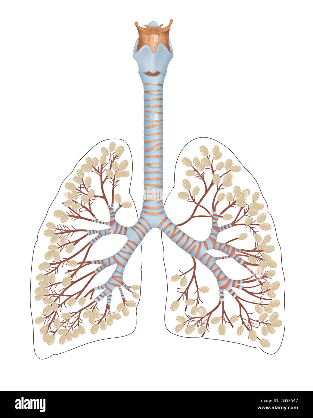
Causes of opacities other than infection. Über 7 millionen englischsprachige bücher. Neurofibromas are a type of noncancerous neoplasm. A neoplasm is an abnormal growth of cells in the lung. What is a soft tissue pulmonary nodule in lung. The alveoli are held open by the transpulmonary pressure, or prestress, which is balanced by tissues forces and alveolar surface film forces. The other main type of liver. The liver parenchyma is the functional tissue of the organ made up of around 80% of the liver volume as hepatocytes. The most prominent structure in this region is the alveolus (figure 1). Lung parenchyma is the substance of the lung that is involved with gas exchange and includes the pulmonary alveoli and respiratory bronchioles, though some authors include only the alveoli. The lung parenchyma is further subdivided into lobes and segments. Analysis of multiple lung parenchymal abnormalities on hrct is a real diagnostic challenge. These abnormalities may be due to a disease of the pulmonary interstitial tissue, the bronchial tree, the cardiovascular system or to abnormal alveolar filling with fluid, blood, cells or tumor, several of these etiologies possibly being concomitant.
The mr imaging appearance of lung parenchyma was investigated with a pulse sequence that offers some solutions to these problems. The video will describe difference between lung parenchyma and lung interstitium. These abnormalities may be due to a disease of the pulmonary interstitial tissue, the bronchial tree, the cardiovascular system or to abnormal alveolar filling with fluid, blood, cells or tumor, several of these etiologies possibly being concomitant. Analysis of multiple lung parenchymal abnormalities on hrct is a real diagnostic challenge. Large holes in the lungs:
Image Of Sarcoidosis Lung Parenchymal Disease from www.meddean.luc.edu Neurofibromas are a type of noncancerous neoplasm. Lung parenchyma is the medical term used to describe the actual functioning parts of a human or animal lung. What is a mural nodule. The other main type of liver. Diffuse parenchymal lung diseases are disorders that affect the interstitial of the lungthe area around the lung's air sacs. Über 7 millionen englischsprachige bücher. These components are not homogeneously distributed over the lung and the relative proportion is continuously changing in function of normal physiological events. In the original analysis, the lung parenchyma was analyzed with a zero boundary condition for the pressure function.
Lung nodules are usually about 0.2 inch (5 millimeters) to 1.2 inches (30 millimeters) in size.
This study found that in a copd cohort changes in lung parenchyma thought to represent interstitial lung disease abnormalities was prevalent (about one out of every 12 hrct scans) and was associated with reduced total lung capacity and a lesser amount of emphysema 77 . Talk to a doctor now. It includes the alveolar walls as well as the blood vessels and the bronchi. The most prominent structure in this region is the alveolus (figure 1). Recent reports have underlined the role of the t lymphocyte as a potentially important factor in the inflammatory process leading to copd. The more cigarettes you smoke per day and the earlier you started smoking, the greater your risk of lung cancer. The most accurate way to determine if a lung disease affects this part of the lung is with a surgical biopsy. Each alveolus in the lung parenchyma opens directly into an alveolar duct or occasionally, in a limited number of species, into a respiratory bronchiole. Über 7 millionen englischsprachige bücher. Lung cyst d is a slow growing lesion usually found incidentally. These components are not homogeneously distributed over the lung and the relative proportion is continuously changing in function of normal physiological events. Brody, md cincinnati children's hospital. He pulmonary parenchyma is the portion of the lung involved in the hematosis or gas transfer.
Diffuse parenchymal lung diseases are disorders that affect the interstitial of the lungthe area around the lung's air sacs. Brody, md cincinnati children's hospital. Pulmonary interstitium is a collection of support tissues within the lung that includes the alveolar epithelium, pulmonary capillary endothelium, basement membrane, perivascular and perilymphatic tissues. Please see disclaimer on my website www.academyofprofessionals.com Lung parenchyma what is lung parenchyma lung parenchyma
Alveoli Of Lungs High Resolution Stock Photography And Images Alamy from c8.alamy.com Areas of consolidation may be patchy, and referred to as bronchopneumonia, or confined to discrete areas of the lung, forming lobar pneumonia. If lung tissue is obtained, however, there is histologic disease in almost all patients, including those who have Interstitial lung disease describes a large group of lung disorders which cause progressive scarring of lung tissue, according to mayo clinic. Lung cyst d is a slow growing lesion usually found incidentally. In interstitial lung disease, some diseases affect all zones while. The liver parenchyma is the functional tissue of the organ made up of around 80% of the liver volume as hepatocytes. Lung parenchyma is the medical term used to describe the actual functioning parts of a human or animal lung. Lung parenchyma is normally considered to be isotropic, that is, its properties do not depend upon specific preferential directions.
The lung parenchyma is further subdivided into lobes and segments.
Recent reports have underlined the role of the t lymphocyte as a potentially important factor in the inflammatory process leading to copd. It includes the alveolar walls as well as the blood vessels and the bronchi. Although this situation may sometimes be approximated in a real lung at low stress, in general there is a finite load imposed by the tissue of the boundary. Über 7 millionen englischsprachige bücher. Interstitial lung disease describes a large group of lung disorders which cause progressive scarring of lung tissue, according to mayo clinic. The lung parenchyma is further subdivided into lobes and segments. Holes in the lungs, known as bullae, can. Magnetic resonance (mr) imaging of lung parenchyma is limited by the low proton density and short t2 in the lung as well as the effects of susceptibility and motion. The other main type of liver. This includes alveoli, alveolar conduits, and respiratory bronchioles. Lung cyst d is a slow growing lesion usually found incidentally. Over time, a granuloma can calcify or harden in the lung, causing a noncancerous lung nodule. Lung parenchyma what is lung parenchyma lung parenchyma


0 Komentar